La Chaîne du Vivant - Chroniques de Biologie d'Yves Muller
By Yves Muller

La Chaîne du Vivant - Chroniques de Biologie d'Yves MullerJun 30, 2021
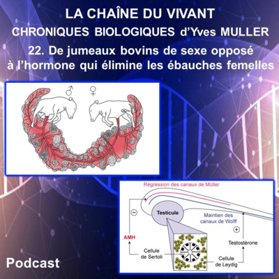
22. De jumeaux bovins de sexe opposé à l’hormone qui élimine les ébauches femelles
Dans cette chronique, vous croiserez 2 faux jumeaux bovins de sexe opposé, des lapins géants des Flandres, une lapine blanche baptisée Céleste et une famille dont les 3 fils possèdent un utérus. Ces rencontres vous permettront de découvrir une étonnante histoire scientifique qui a conduit à la caractérisation d’une nouvelle hormone impliquée dans la différenciation sexuelle. Quel que soit son sexe génétique, l’embryon de Mammifère possède 2 canaux de Wolff et 2 canaux de Müller. Si l’embryon est de sexe génétique mâle, les 2 canaux de Müller vont régresser tandis que les 2 canaux de Wolff seront maintenus et formeront les voies génitales mâles sous l’influence de la testostérone produite par le testicule fœtal. Si l’embryon est de sexe génétique femelle, les 2 canaux de Wolff vont régresser tandis que les 2 canaux de Müller seront maintenus et ils formeront les voies génitales femelles. Cette chronique présente les travaux d’Alfred Jost publiés en 1947 et qui lui ont permis d’affirmer que la testostérone n’était pas la seule hormone produite par le testicule fœtal, mais qu’il devait exister une autre hormone responsable de la régression des canaux de Müller. Puis vous verrez que le travail acharné et passionné de Nathalie Josso lui a permis en 1984 d’isoler cette nouvelle hormone testiculaire, qu’elle a nommé Hormone anti-Müllerienne ou AMH. La suite de cette histoire scientifique révèlera que l’AMH est produite aussi par l’ovaire et le dosage sanguin de l’AMH chez la femme permet aujourd’hui d’évaluer les chances de fertilité féminine. Vous verrez aussi que la découverte de l’AMH a permis de comprendre pourquoi dans certaines familles, les garçons ont un utérus.
Errata : - à 5'02, l'année est 1780 et non 1880.
- à 13'09, Leydig a découvert les cellules de Leydig en 1857, donc avant la découverte par Sertoli en 1865 des cellules de Sertoli.
Autuer
Références :
- Keller K et Tandler J (1916) The behaviour of the chorion in twin pregnancies of cattle - Wien. Tierärztl. Monatsschr ; 3:513-516.
- Lillie FR (1917) Ther freemartin, a study of the action of sex hormones in foetal life of cattle - J. Exp. Zool. ; 23:371-452.
- Picon R (1969) Action du testicule foetal sur le développement in vitro des canaux de Müller chez le rat – Arch. Anat. Micros. Morph. Exp ; 58:1-19.
- Jost A et coll. (1972) Becoming a male - Adv. Biosci ; 10:3-13.
- Josso N (1973) In vitro synthesis of Müllerian-inhibiting hormone by seminiferous tubules isolated from the calf fetal testis – Endocrinology ; 93:829-834.
- Picard JY et Josso N (1984) Purification of testicular anti-Müllerian hormone allowing direct visualization of the pure glycoprotein and determination of yield and purification factor -Molecular and Cellular Endocrinology ; 34:23-29.
- Guerrier D et coll. (1989) The persistent Müllerian duct syndrome: a molecular approach - Journal of Clinical Endocrinology and Metabolism ; 68:46-52.
- Knebelmann B et coll. (1991) Anti-Müllerian hormone Bruxelles : a nonsense mutation associated with the persistent Müllerian duct syndrome – PNAS USA ; 88:3767-3771.
- di Clemente N et coll. (1994) Cloning, expression, and alternative splicing of the receptor for anti-Müllerian hormone – Molecular Endocrinology ; 8:1006-1020.
- Racine C et coll. (1998) Receptors for anti-mullerian hormone on Leydig cells are responsible for its effects on steroidogenesis and cell differentiation. PNAS USA ; Proceedings of the National Academy of Sciences (USA) ; 95:594-599.
- Durlinger AL et coll. (1999) Control of primordial follicle recruitment by anti- Mullerian hormone in the mouse ovary ; Endocrinology ; 140:5789-5796.
- van Rooij IAJ, Themmen APN et coll. (2002) Serum anti-Müllerian hormone levels: A novel measure of ovarian reserve - Human Reproduction ; 17:3065-3071.
- Josso N (2017) Le Sexe des Anges : Une histoire d'hormones - EDP Sciences
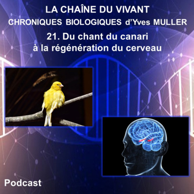
21. Du chant du canari à la régénération du cerveau (nouvelle version)
Cette chronique démarre par le chant de séduction qu’émet le canari mâle au printemps et que cet oiseau chanteur va modifier chaque année. Vous croiserez ensuite le Dieu Pan jouant de la flûte, des mésanges cachant des graines, des souris qui apprennent et mémorisent, des chauffeurs de taxi de la ville de Londres et des essais nucléaires pendant la guerre froide. Cette chronique vous conduira à découvrir que le cerveau des oiseaux et des mammifères adultes est pourvu d’une capacité de régénération. Et peut-être parviendrons-nous un jour à stimuler la production de neurones dans notre cerveau pour faire face à des lésions cérébrales ou des maladies neurodégénératives.
Auteur : Yves Muller - Professeur Agrégé de classe exceptionnelle - Docteur en Neurosciences - Université de Montpellier
21. Biological Chronicles - From the song of the canary to the regeneration of the brain
Our chronicle begins with the seduction song the male canary emits in spring and which this songbird will modify each year. We will then encounter the God Pan playing the flute, chickadees hiding seeds, mice with learning and memorizing capabilities, London taxi drivers and nuclear tests during the Cold War. Our chronicle leads us to then to the discovery that the brains of adult birds and mammals have a capacity for regeneration. And maybe we’ll one day be able to stimulate the production of neurons in our brain to deal with brain damage or neurodegenerative diseases. From a fly with legs on its head to the discovery architectural genes
Author : Yves Muller – Associate Professor at Montpellier University
Références :
- Messier B, Leblond CP, Smart I (1958) Presence of DNA synthesis and mitosis in the brain of young adult mice - Experimental Cell Research ; 14:224–6
- Altman J et Das GD (1965) Autoradiographic and histological evidence of postnatal hippocampal neurogenesis in rats - Journal of Comparative Neurology ; 124:319-35
- Nottebohm F, Stokes TM, Leonard CM (1976) Central control of song in the canary, Serinus canaria - Journal of Comparative Neurology ; 165: 457–86
- Nottebohm F (1981) A brain for all seasons: cyclical anatomical changes in song control nuclei of the canary brain - Science ; 214:1368-70
- Paton JA et Nottebohm FN (1984) Neurons generated in the adult brain are recruited into functional circuits – Science ; 225:1046-8
- Nottebohm F (1989) From Bird Song to Neurogenesis - Scientific American ; 260:74-9
- Alvarez-Buylla A, Theelen M et Nottebohm F (1990) Proliferation “hot spots” in adult avian ventricular zone reveal radial cell division – Neuron ; 5:101–9
- Kirn J, O’Loughlin B et coll. (1994) Cell death and neuronal recruitment in the high vocal center of adult male canaries are temporally related to changes in song - PNAS USA ; 91:7844-8
- Barena A et Nottebohm F (1994) Seasonal recruitment of hippocampal neurons in adult free-ranging black-capped chickadees - PNAS USA ; 91:11217-21
- Eriksson PS, Perfilieva E et coll. (1998) Neurogenesis in the adult human hippocampus - Nature Medicine ; 4:1313-7
- Taupin P (2005) Neurogenesis in the pathologies of the nervous system - Medecine Sciences 21:711-4
- Nottebohn F (2005) The neural basis of birdsong – PloS Biology ; 3:e164,759-61
- Rapanelli M, Frick L et Zanutto B (2011) Learning an operant conditioning task differentially induces gliogenesi in the medial prefrontal cortex and neurogenesis in the hippocampus - PLoS One 2011 ; 6:e14713, 1-12
- Woollett K et Maguire E (2011) Acquiring “the Knowledge” of London's layout drives structural brain changes - Current Biology ; 21:2109-14
- Bergmann O, Liebl J et coll. (2012) The age of olfactory bulb neurons in humans – Neuron ; 74:634-9
- Alonso M, Lepousez G et coll. (2012) Activation of adult-born neurons facilitates learning and memory - Nature Neuroscience ; 15:897-904
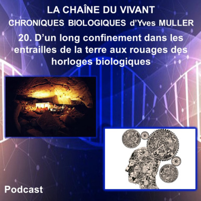
20. D’un long confinement dans les entrailles de la terre aux rouages des horloges biologiques
Cette 20ème chronique démarre en été 1962, avec un spéléologue dénommé Michel Siffre, qui s'enfonce dans le noir du gouffre de Scarasson à la frontière franco-italienne. Et il va passer 60 jours seul dans les entrailles de la terre. Avec cette expérience d'isolement hors du temps, une nouvelle science vient de naître, la chronobiologie. Cette chronique présente ensuite les travaux qui ont permis le décryptage des mécanismes moléculaires de l’horloge biologique d’une petite mouche et qui ont valu à leurs auteurs le prix Nobel 2017 de physiologie. Les rouages des horloges biologiques des Mammifères sont ensuite abordés par comparaison avec ceux de la mouche, mettant en évidence l’existence d’un modèle de fonctionnement commun, mais faisant aussi apparaître des différences majeures, notamment dans le mode d’action de la lumière dans la synchronisation des horloges.
Auteur : Yves Muller – Professeur Agrégé de classe exceptionnelle - Docteur en Neurosciences - Université de Montpellier
Biological Chronicles - From a long confinement in the bowels of the earth to the cogs of our biological clocks
Our 20th chronicle begins in summer 1962, with a speleologist named Michel Siffre, descending into the dark of the Scarasson abyss on the Franco-Italian border. He will spend 60 days alone in the bowels of the earth. Through this experience of timeless isolation, a new science is born, chronobiology. Our chronicle then presents the work that allowed for the decoding of the molecular mechanisms of the biological clock of a small fly and that won their authors the 2017 Nobel Prize in physiology. We then compare the workings of the biological clocks in Mammals with those of the fly, highlighting the existence of a common operating model, but also major differences, notably in the mode of action of light in clock synchronization.
Références :
De Mairan J. (1729) Observation botanique. Hist Acad Roy Sci ; 35-36
Michel Siffre (1963) Hors du temps. Julliard
Konopka RJ, Benzer S. (1971) Clock mutants of Drosophila melanogaster. Proc Natl Acad Sci USA ; 68 : 2112–6.
Reddy P, Zehring WA, Wheeler DA, et al. (1984) Molecular analysis of the period locus in Drosophila melanogaster and identification of a transcript involved in biological rhythms. Cell ; 38 : 701-10.
Crews ST, Thomas JB, Goodman CS. (1988) The Drosophila single-minded gene encodes a nuclear protein with sequence similarity to the per gene product. Cell ; 52 : 143-51.
Hardin PE, Hall JC, Rosbash M. (1990) Feedback of the Drosophila period gene product on circadian cycling of its messenger RNA levels. Nature ; 343 : 536-40.
Hardin PE, Hall JC, Rosbash M. (1992) Circadian oscillations in period gene mRNA levels are transcriptionally regulated. Proc Natl Acad Sci USA ; 89 :11711-5
Zeng H, Hardin PE, Rosbash M. (1994) Constitutive overexpression of the Drosophila period protein inhibits period mRNA cycling. EMBO J ; 13 : 3590-8
Sehgal A, Price JL, Man B, Young MW. (1994) Loss of circadian behavioral rhythms and per RNA oscillations in the Drosophila mutant timeless. Science 263 : 1603-6
Vosshall LB, Price JL, Sehgal A, Saez L, Young MW. (1994) Block in nuclear localization of period protein by a second clock mutation, timeless. Science ; 263 : 1606-9
Vitaterna MH, King DP, Chang AM et al. (1994) Mutagenesis and mapping of a mouse gene, Clock, essential for circadian behavior. Science ; 264 :719-25
Gekakis N, Saez L, Delahaye-Brown AM et al. (1995) Isolation of timeless by PER protein interaction: defective interaction between timeless protein and long-period mutant PERL. Science ; 270 : 811-5
Price JL, Dembinska ME, Young MW, Rosbash M. (1995) Suppression of PERIOD protein abundance and circadian cycling by the Drosophila clock mutation timeless. EMBO J ; 14 : 4044-9
Ameisen JC (2017) Les horloges circadiennes – Sur les épaules de Darwin - France Inter
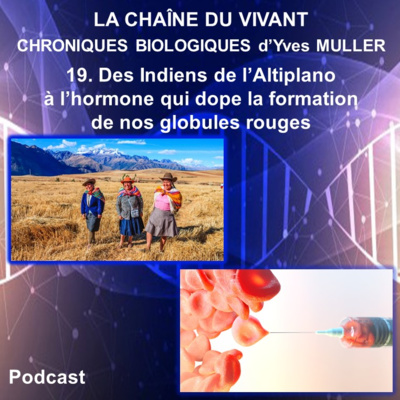
19. Des Indiens de l’Altiplano à l’hormone qui dope la production de nos globules rouges
Cette 19ème chronique démarre sur l’Altiplano, où vivent des populations andines adaptées à la haute altitude, et donc exposées à une hypoxie chronique. Le sang des andins contient un nombre de globules rouges supérieur à celui des populations qui vivent au niveau de la mer. Cette chronique explique ensuite comment le manque d’oxygène en altitude conduit à la production par les reins de l’érythropoïétine (ou EPO), une hormone de nature protéique qui stimule la production des globules rouges. Les mécanismes moléculaires qui permettent à l’hypoxie d’activer l’expression de gènes, tel que le gène de l’EPO, sont présentés à partir des résultats des 3 chercheurs qui ont obtenu en 2019 le Prix Nobel de Physiologie ou de Médecine. Et la compréhension de ces mécanismes qui permettent aux cellules de s’adapter aux apports variables d’oxygène offrent de nouvelles perspectives pour lutter contre l’anémie et le cancer.
Auteur : Yves Muller – Professeur Agrégé de classe exceptionnelle - Docteur en Neurosciences - Université de Montpellier
Biological Chronicles - From the Altiplano Indians to the hormone that boosts the production of red blood cells
Our 19th chronicle begins on the Altiplano, hope to Andean populations who have adapted to high altitude, and are therefore exposed to chronic hypoxia. Andean blood contains a higher number of red blood cells than that of people living at sea level. Our chronicle then explains how lack of oxygen at high altitudes leads the kidneys to produce erythropoietin (or EPO), a protein hormone that stimulates the production of red blood cells. The molecular mechanisms that allow hypoxia to activate the expression of genes, such as the EPO gene, are presented based on the results of 3 researchers who won the 2019 Nobel Prize in Physiology or Medicine. Understanding the mechanisms which allow cells to adapt to variable oxygen supplies offers new perspectives in the fight against anemia and cancer.
Références :
- Semenza, G.L, Nejfelt, M.K., Chi, S.M. & Antonarakis, S.E. (1991). Hypoxia-inducible nuclear factors bind to an enhancer element located 3’ to the human erythropoietin gene. Proc Natl Acad Sci USA, 88, 5680-5684
- Wang, G.L., Jiang, B.-H., Rue, E.A. & Semenza, G.L. (1995). Hypoxia-inducible factor 1 is a basic-helix-loop-helix-PAS heterodimer regulated by cellular O2 tension. Proc Natl Acad Sci USA, 92, 5510-5514
- Maxwell, P.H., Wiesener, M.S., Chang, G.-W., Clifford, S.C., Vaux, E.C., Cockman, M.E., Wykoff, C.C., Pugh, C.W., Maher, E.R. & Ratcliffe, P.J. (1999). The tumour suppressor protein VHL targets hypoxia-inducible factors for oxygen-dependent proteolysis. Nature, 399, 271-275
- Ivan, M., Kondo, K., Yang, H., Kim, W., Valiando, J., Ohh, M., Salic, A., Asara, J.M., Lane, W.S. & Kaelin Jr., W.G. (2001) HIFatargeted for VHL-mediated destruction by proline hydroxylation: Implications for O2 sensing. Science, 292, 464-468
- Jaakkola, P., Mole, D.R., Tian, Y.-M., Wilson, M.I., Gielbert, J., Gaskell, S.J., von Kriegsheim, A., Heberstreit, H.F., Mukherji, M., Schofield, C.J., Maxwell, P.H., Pugh, C.W. & Ratcliffe, P.J. (2001). Targeting of HIF-α to the von Hippel-Lindau ubiquitylation complex by O2-regulated prolyl hydroxylation. Science, 292, 468-472
- A. Bigham A, M. Bauchet et al. (2010) - Identifying signatures of natural selection in Tibetan and Andean populations using dense genome scan data - PLOS Genetics 6 (9)
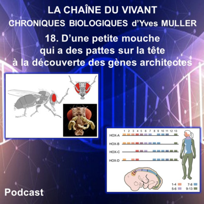
18. D’une petite mouche qui a des pattes sur la tête à la découverte des gènes architectes
Cette 18ème chronique démarre par une étrange petite mouche pourvue de pattes sur la tête à la place des antennes. Il s’agit d’un mutant de drosophile appelé Antennapedia. Un autre mutant de drosophile, appelé Ultrabithorax, présente 2 paires d’ailes et non une seule. Et l'étude approfondie de tels mutants va permettre de découvrir les gènes homéotiques Hox, dont le fonctionnement détermine le plan du corps de la drosophile. La découverte de ces gènes architectes chez la petite mouche va permettre d’identifier chez les Vertébrés des gènes homéotiques Hox, apparentés à ceux de la drosophile, et qui déterminent le plan du corps des Vertébrés ainsi que la formation de leurs membres. Les gènes Hox, présents chez tous les animaux pluricellulaires, codent pour des facteurs de transcription à homéodomaine qui contrôlent le développement en régulant des réseaux de gènes impliqués dans la structuration du corps. Et ces gènes Hox architectes du plan du corps ont dû jouer un rôle fondamental dans l'évolution et la diversification des animaux segmentés.
Auteur : Yves Muller – Professeur Agrégé de classe exceptionnelle - Docteur en Neurosciences - Université de Montpellier
Biological Chronicles - From a fly with legs on its head to the discovery architectural genes
Our 18th chronicle begins with a strange little fly with legs on its head instead of antennae. It is a Drosophila mutant known as Antennapedia. Another Drosophila mutant, Ultrabithorax, has 2 pairs of wings instead of one. The in-depth study of such mutants will bring about the discovery of homeotic Hox genes, whose general function is to determine the body plan of Drosophila. The discovery of these architectural genes in the small fly will make it possible to identify homeotic Hox genes in vertebrates related to Drosophila as well. The Hox genes determine the body plan of vertebrates and also the formation of their members. The Hox genes, present in all multicellular animals, encode homeodomain transcription factor proteins which control development by regulating gene networks involved in the structural organization of the body. These Hox genes, architects of the body plan, must have played a fundamental role in the evolution and diversification of segmented animals.
Références :
- Bateson W (1894) Materials for the study of variation, treated with especial regard to dis-continuity in the origin of species – Londres, MacMillan and Co
- Lewis EB (1978) A gene complex controlling segmentation in Drosophila – Nature ; 76:565-70
- Nüsslein-Volhard C et Wieschaus E (1980) Mutations affecting segment number and polarity in Drosophila - Nature ; 287:795-801
- Bender W et coll. (1983) Molecular genetics of the bithorax complex in Drosophila melanogaster – Science ; 221:23-9
- Scott MP et Weiner AJ (1984) Structural relationships among genes that control development: sequence homology between the antennapedia, ultrabithorax, and fushi tarazu loci of Drosophila – PNAS USA ; 81:4115-9
- McGinnis W et coll. (1984) A conserved DNA sequence in homoeotic genes of the Drosophila Antennapedia and bithorax complexes - Nature ; 308:428–33
- Denis Duboule (2007) The rise and fall of Hox gene clusters - Development ; 134:2560
- Wellik DM (2009) Hox genes and vertebrate axial pattern - Curr Top Dev Biol ; 88:257-78
- Mallo M et coll. (2010) Hox genes and regional patterning of the vertebrate body plan - Dev Biol ; 344:7-15
- Gaunt SJ (2018) Hox cluster genes and collinearities throughout the tree of animal life - The International Journal of Developmental Biology ; 62: 673–83
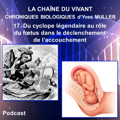
17. Du cyclope légendaire au rôle du fœtus dans le déclenchement de l’accouchement
Cette 17ème chronique biologique démarre par une première histoire contée par Homère dans l’Iliade et qui oppose le héros Ulysse au cyclope Polyphème. Et dans cette histoire mythologique, ce sont des moutons qui vont aider Ulysse à s’échapper de l’antre du cyclope. Puis cette chronique raconte une seconde histoire, véridique cette fois, dans laquelle on parle aussi de moutons et de cyclopes, mais aussi de la petite mouche du vinaigre puis de Sonic le hérisson, personnage célèbre de jeux vidéo. Cette seconde histoire nous amène à décrire les mécanismes qui permettent à la brebis de mettre bas, en montrant que c’est le fœtus lui-même qui va déclencher la mise bas. Puis les mécanismes déclenchant l’accouchement chez la femme sont également abordés, par comparaison avec la brebis. Cette chronique nous conduit donc étonnamment du cyclope au mécanismes de l’accouchement, mais elle nous amène de façon tout aussi surprenante à de nouvelles pistes médicamenteuses pour lutter contre certains cancers.
Auteur : Yves Muller – Professeur Agrégé de classe exceptionnelle - Docteur en Neurosciences - Université de Montpellier
Biological Chronicles - From the legendary cyclops to the role of the fetus in the onset of childbirth.
This 17th biological chronicle begins with a story told by Homer in the Iliad which pits the hero Ulysses against Cyclops Polyphemus. In this mythological story, Ulysses is helped to escape from the cave of the Cyclops by sheep. Then, our chronicle will tell a second story, true this time, which is also about sheep and cyclops, but also about tiny vinegar flies and Sonic the hedgehog (famous video game character). This second story leads us to the mechanisms that allow sheep to give birth, showing that it is the fetus itself that triggers the start of labor. The mechanisms triggering childbirth in women are also discussed, in comparison to sheep. This chronicle thus brings us surprisingly from the cyclops to the mechanisms of childbirth, but it brings us just as surprisingly to new treatment possibilities in the fight against certain cancers.
Références :
I only have eye for ewe: the discovery of cyclopamine and development of Hedgehog pathway-targeting drugs – Chen JK - Nat Prod Rep. 2016 May 4;33(5):595-601.
Cyclopamine and Hedgehog Signaling: Chemistry, Biology, Medical Perspectives - Philipp Heretsch Lito Tzagkaroulaki, Athanassios Giannis – 2010 - Angewandte Chemie International Edition/Volume 49, Issue 20
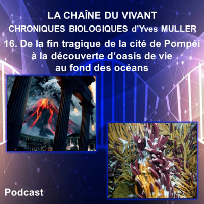
16. De la fin tragique de la cité de Pompéi à la découverte d’oasis de vie au fond des océans
Cette 16ème chronique démarre en l’an 79 après Jésus-Christ, au moment où le Vésuve entre en éruption et détruit en seulement 2 jours la cité romaine de Pompéi. Puis, cette chronique se poursuit 20 siècles plus tard avec l’extraordinaire découverte en 1977 d’étonnantes communautés animales qui se développent dans les profondeurs océaniques, là où les sources hydrothermales associées aux dorsales océaniques créent un environnement que l’on pourrait croire très défavorable à la vie. Parmi les surprenants animaux rencontrés dans ces environnement extrêmes est abordée une espèce emblématique de ces milieux, un ver tubicole géant appelé Riftia pachyptila. Et avec la description de cet étrange ver dépourvu de système digestif est présentée la symbiose entre ce ver et les bactéries chimiosynthétiques qu’il héberge dans son corps. Une telle symbiose se retrouve dans d’autres animaux de ces sites hydrothermaux, tel que le grand Bivalve Calyptogena magnifica. Et près des cheminées hydrothermales ont aussi été identifiés des vers surprenant, appelés Vers de Pompéi (ou Alvinella pompejana) et qui présentent une tolérance exceptionnelle à la chaleur. Sont ensuite abordés les réseaux trophiques de ces écosystèmes qui reposent sur les bactéries chimiosynthétiques. Puis l’intérêt que présente pour l’homme l’étude de ces écosystèmes hydrothermaux océaniques est abordé en fin de chronique.
Auteur : Yves Muller – Professeur Agrégé Université Montpellier
Biological Chronicles - From the tragic end of the city of Pompeii to the discovery of oases of life on the ocean floor.
This 16th chronicle begins in the year 79 AD, when the eruption of Mt. Vesuvius destroys the Roman city of Pompeii in just 2 days. Our chronicle picks up 20 centuries later in 1977 with the extraordinary discovery of amazing animal communities in the ocean depths, where the hydrothermal vents associated with the ocean ridges create an environment that one might think quite unfavorable to life. Among the surprising animals encountered in these extreme environments is a giant tubeworm called Riftia pachyptila. This strange worm, devoid of a digestive system shows us the symbiosis between itself and the chemosynthetic bacteria that it hosts in its body. Such a symbiosis is found in other animals of these hydrothermal sites, such as the great Bivalve Calyptogena magnifica. Near the hydrothermal vents other surprising worms have also been identified, the Pompeii worm (or Alvinella pompejana) which have an exceptional tolerance to heat. We’ll then move on to the trophic networks of these ecosystems, which are based on chemosynthetic bacteria. To finish this chronicle, we’ll discuss the interest of humans in the study of these oceanic hydrothermal ecosystems.
Références :
Pline le Jeune (environ 100 ap. J.-C.) – Lettres de Pline, livre I à X – Traduction d’Annette Flobert – Editions Flammarion (2002)
Corliss J, Dymond J et coll. (1979) Submarine Thermal Springs on the Galápagos Rift - Science ; 4385:1073-1083
Desbruyères D et Laubier L (1980) Alvinella pompejana gen.sp. nov., Ampharetidae aberrant des sources hydrothermales de la ride Est-Pacifique - Oceanologica Acta ; 3:267-274
Childress J, Fisher CR (1992). The biology of hydrothermal vent animals: physiology, biochemistry, and autotrophic symbioses - Oceanography and Marine Biology: An Annual Review ; 30:337-441
Laubier L (2008) Ténèbres océanes. Le triomphe de la vie dans les abysses - Editions Buchet Chastel
Hekinian R et Binard N (2008) Le feu des abysses – Editions Quæ
Lallier F et coll. (2009) La chimiosynthèse apprivoisée : des modèles venus de l’océan profond - Biofutur ; 299 :45-4
Duperron S (2017) Les symbioses microbiennes, associations au cœur du vivant - Editions ISTE
Karsenti E (2018) Aux sources de la vie – Editions Flammarion
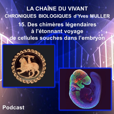
15. Des chimères légendaires à l’étonnant voyage de cellules souches dans l’embryon
Ce sont des créatures légendaires issues de la mythologie grecque qui ouvrent cette 15ème chronique. On y découvre notamment la Chimère dont le corps est un mélange de lion, de chèvre et de serpent. On y rencontre aussi un cheval ailé dénommé Pégase, des centaures, le Sphinx et des sirènes. Puis cette chronique présente des animaux chimériques bien réels cette fois et que des biologistes ont construits dans le but d’étudier certains mécanismes du développement. Ce sont des chimères embryonnaires, obtenues chez les amphibiens, puis chez les oiseaux, qui sont abordées dans cette chronique. Le premier exemple de chimère embryonnaire qui est présenté ici est issu des travaux d’embryologie expérimentale réalisés en 1924 par Hans Spemann et Hilde Mangold à partir de 2 espèces différentes de tritons. Et ces travaux ont permis de mettre en évidence l’existence d’un processus d’induction dans l’embryon. Le second exemple est celui des embryons chimériques caille-poulet obtenus par Nicole Le Douarin à partir des années 1970 et qui ont notamment révélé le voyage et le devenir des cellules des crêtes neurales dans l’embryon. Vous découvrirez ainsi que la crête neurale céphalique a joué un rôle clé dans l’histoire évolutive des Vertébrés et qu’elle a largement contribué à spécifier l’identité de leurs visages. Et l’utilisation des cellules des crêtes neurales ou de leurs dérivés offre aujourd'hui de nouveaux espoirs en médecine régénérative.
Auteur : Yves Muller – Professeur Agrégé Université Montpellier
Biological Chronicles - From legendary chimeras to the astonishing journey of stem cells in the embryo
Our 15th chronicle opens with legendary creatures from Greek mythology, in particular, the Chimera, part lion, part goat, part serpent. We will also meet a winged horse called Pegasus, centaurs, the Sphinx and mermaids. Our chronicle then moved on to fanciful animals, this time very real, that biologists have built in order to study certain mechanisms of development. These are embryonic chimeras, obtained from amphibians, then from birds. The first example of an embryonic chimera which is presented here comes from experimental embryology work carried out in 1924 by Hans Spemann and Hilde Mangold from 2 different species of newts. Their work has made it possible to highlight the existence of an embryonic induction process. Our second example is of chimeric quail-chicken embryos obtained by Nicole Le Douarin in the 1970s and which in particular revealed the journey and fate of neural crest cells in the embryo. We will discover that the cephalic neural crest has played a key role in the evolutionary history of vertebrates as well as a major role in specifying their facial identity. The use of neural crest cells or their derivatives now offers new hope in regenerative medicine.
Références :
- Spemann H et Mangold H (1924) Induction of embryonic primordia by implantation of organizers from a different Species - Archiv Für Mikroskopische Anatomie Und Entwicklungsmechanik ; 100:599-638
- Le Douarin N (1969) Particularités du noyau interphasique chez la Caille japonaise (Coturnix coturnix japonica). Utilisation de ces particularités comme « marquage biologique » dans les recherches sur les interactions tissulaires et les migrations cellulaires au cours de l'ontogenèse - Bull Biol Fr Belg ; 1103:435-52
- Le Douarin N (1973) A biological cell labeling technique and its use in experimental embryology - Developmental Biology ; 30:217-22
- Le Douarin N (1982) The Neural Crest - Cambridge : Cambridge University Press
- Fässler P et Sander K (1996) Hilde Mangold and Spemann's organizer : achievement and tragedy - Development Genes and Evolution ; 205:323-32
- Le Douarin N et Kalcheim C (1999) The Neural Crest. 2nd ed. New York : Cambridge University Press
- Le Douarin N (2000) Des chimères, des clones et des gènes - Editions Odile Jacob
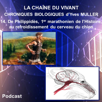
14. De Philippidès, premier marathonien de l’Histoire, au système de refroidissement du cerveau du chien
Cette 14ème chronique démarre il y a plus de 25 siècles avec le célèbre exploit de Philippidès qui courut de Marathon à Athènes pour annoncer la victoire des siens sur les Perses avant de succomber brutalement à son effort. Le cerveau est particulièrement sensible à l’hyperthermie et la mort foudroyante de Philippidès pourrait être due à une hyperthermie cérébrale. Bien qu’il existe chez l’homme des mécanismes de refroidissement du cerveau, ils s’avèrent moins efficaces que ceux qui existent chez certains Mammifères, tels que le chien ou la gazelle. En effet, ces animaux possèdent à la base de leur cerveau un réseau admirable, appelé le plexus carotidien, qui permet lors d’un effort d’éviter l’hyperthermie cérébrale.
Auteur : Yves Muller – Professeur Agrégé de classe exceptionnelle - Docteur en Neurosciences - Université de Montpellier
Errata : à 2.20 , la phrase exacte est : "La réponse faite par les spartiates à Philippidès..." au lieu de "La réponse faite par les perses à Philippidès..."
Biological Chronicles - From Philippides, the first marathon runner in history, to the cooling system of the dog's brain
Our 14th chronicle begins over 25 centuries ago with the famous feat of Philippides who ran from Marathon to Athens to announce his people’s victory over the Persians before succumbing suddenly to his effort. The brain is particularly sensitive to hyperthermia and the devastating death of Philippides could be due to cerebral hyperthermia. Although brain cooling mechanisms exist in humans, they are less effective than those that exist in some mammals, such as dogs and gazelles. Indeed, at the base of the brain these animals have an impressive network called the carotid plexus which prevents cerebral hyperthermia during physical exertion.
Références :
- Hérodote (5ème siècle av. J.-C.) Histoires - Livre VI. Edition Gallimard 1990
- Plutarque (1er - 2e siècle ap. J.-C.) La gloire des Athéniens - Œuvres morales. Edition Les Belles Lettres 1970
- Taylor CR et Lyman CP. (1972) Heat storage in running antelopes: independence of brain and body temperatures. The American Journal of Physiology ; 222(1):114-7
- Baker MA et Chapman LW (1977) Rapid brain cooling in exercising dogs. Science ; 195(4280):781-3
- Baker MA (1979) A brain-cooling system in mammals. Scientific American ; 240(5):130-9
- Germain M, Jobin M, Cabanac M (1987) The effect of face fanning during recovery from exercise hyperthermia. Canadian Journal of Physiology and Pharmacology ; 65:87-91
- Cabanac M et Bonniot-Cabanac M-C (1997) De quoi serait mort le coureur de Marathon ? médecine/sciences ; 13:838-42
- Cavaillon J-M (1998) Diagnostic post-mortem du coureur de marathon : une contre-expertise !!! médecine/sciences ; 14:132
- Heffernan KS (2012) How healthy were the arteries of Phidippides ? Clinical Cardiology ; 35 : 65-8

13. D'un chocolat doux comme un baiser au contrôle de la reproduction des Mammifères
Cette 13ème chronique biologique démarre dans la ville de Hershey aux États-Unis, où l’on trouve une chocolaterie qui fabrique des « Hershey’s Kisses » (baisers d'Hershey). Et à partir de ces chocolats doux comme des baisers, je vous propose un voyage dans l’histoire des sciences à la découverte du gène KISS-1 et des kisspeptines. Cette chronique vous amènera à comprendre les mécanismes de contrôle neuroendocrinien de la reproduction des Mammifères et elle vous montrera que les kisspeptines jouent un rôle le rôle essentiel dans ces mécanismes.
Auteur : Yves Muller – Professeur Agrégé de classe exceptionnelle - Docteur en Neurosciences - Université de Montpellier
Biological Chronicles - From sweet chocolate like a kiss to mammalian reproductive control
This 13th biological chronicle presents the kisspeptin system. The kisspeptin system is believed to be requisite for normal GnRH secretion, serving as a “gatekeeper” of puberty and helping to mediate the effects of sex steroids on GnRH secretion. Kisspeptin was originally called metastin because of its ability to suppress metastatic spread of human melanomas. However, in recognition of its discovery at Pennsylvania State University in Hershey, Pennsylvania, it was later named kisspeptin after Hershey’s chocolate Kisses.
Références :
- Lee JH, Miele ME, Hicks DJ, Phillips KK, Trent JM, Weissman BE, Welch DR (1996). "KiSS-1, a novel human malignant melanoma metastasis-suppressor gene" - Journal of the National Cancer Institute ; 88: 1731–7
- Kotani M, Detheux M, Vandenbogaerde A et al. (2001) The metastasis suppressor gene KiSS-1 encodes kisspeptins, the natural ligands of the orphan G protein-coupled receptor GPR54 - J Biol Chem ; 276:34631–34636
- Ohtaki T, Shintani1 Y, Honda S et al. (2001) Metastasis suppressor gene KiSS-1 encodes peptide ligand of a G-protein-coupled receptor - Nature ; 411:613–617
- Han SK, Gottsch ML, Lee KJ et al. (2005) Activation of gonadotropin-releasing hormone neurons by kisspeptin as a neuroendocrine switch for the onset of puberty - J Neurosci ; 7:11349–11356
- Dhillo WS, Chaudhri OB, Thompson EL et al. (2007) Kisspeptin-54 stimulates gonadotropin release most potently during the preovulatory phase of the menstrual cycle - J Clin Endocrinol Metab ; 92:3958–3966
- George JT, Veldhuis JD, Roseweir AK et al. (2011) Kisspeptin is a potent stimulator of LH and increases pulse frequency in men - J Clin Endocrinol Metab ; 96:1228–1236
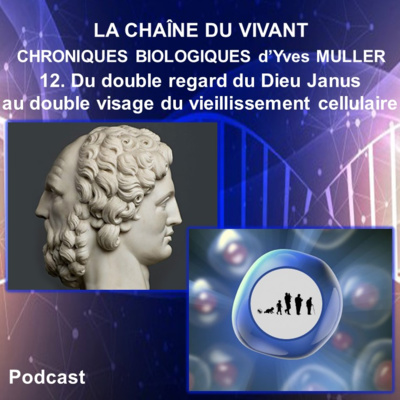
12. Du double regard du Dieu Janus au double visage de la sénescence cellulaire
C’est le Dieu Janus de la mythologie romaine qui ouvre cette 12ème chronique biologique. Janus est le Dieu des portes et des transitions, l’Ouvreur et le Fermeur, l’éternel Passeur du monde. On le représente souvent avec un double visage qui lui permet de voir à la fois derrière et devant lui. Après cette ouverture mythologique, la chronique aborde une destinée particulière de nos cellules appelée la sénescence cellulaire. Elle correspond à un vieillissement des cellules, devenues alors incapables de division. Cette chronique présente les signaux qui induisent la sénescence des cellules, comme l’érosion des télomères, des stress oxydants ou génotoxiques et l’activation d’oncogènes. Puis sont décrites les caractéristiques morphologiques et fonctionnelles de la cellule sénescente. Cette chronique montre que la sénescence cellulaire, comme le Dieu Janus, a un double visage. Elle a un visage avenant et bénéfique puisqu’elle favorise la cicatrisation et nous protège du cancer. Mais la sénescence cellulaire offre un second visage, sombre et maléfique car elle contribue au vieillissement de notre corps et aux pathologies associées à l’âge. Des laboratoires développent actuellement des substances sénolytiques capables de détruire sélectivement les cellules sénescentes. On peut rêver qu’un jour prochain, l’utilisation de telles substances nous permettra de prolonger notre espérance de vie en bonne santé et de poursuivre plus longtemps notre voyage dans le temps.
Auteur : Yves Muller - Professeur Agrégé de classe exceptionnelle - Docteur en Neurosciences - Université de Montpellier
12. Biological Chronicles - From the two faces of the God Janus to the two faces of cellular aging
God Janus of Roman mythology opens our 12th biological chronicle. Janus is the God of doorways and transitions, the Opener and the Closer, the eternal Ferryman of the world. We often represent him as having two faces which allows him to see both behind and ahead. After this mythological opening, our chronicle addresses a particular destiny of our cells called cellular senescence. That being an aging of the cells, which then become incapable of division. We then discuss the signals that induce cellular senescence, such as the erosion of telomeres, oxidative or genotoxic stresses and the activation of oncogenes. We then move on to the morphological and functional characteristics of a senescent cell. This chronicle shows that cellular senescence, like the God Janus, has two faces. The first is attractive and beneficial since it promotes healing and protects us from cancer. But cellular senescence offers a second, darker face because it contributes to the aging of our bodies and to the pathologies associated with age. Laboratories are currently developing senolytic substances capable of selectively destroying senescent cells. We can hope that one day soon, the use of such substances will allow us to live longer, healthier lives and to continue our travels farther into the future.
Références :
- Hayflick L, Moorhead P (1961) The serial cultivation of human diploid cell strains. Experimental Cell Research ; 25:585-621
- Ameisen JC (2017) – Le navire de Thésée – Sur les épaules de Darwin – Emission de radio sur France Inter
- Bourgeois B et Madle T (2018) Regulation of cellular senescence via the FOXO4-p53 - FEBS Letters ; 592:2083–2097
- Xu M, Pirtskhalava T, Farr JN, et al. (2018) Senolytics improve physical function and increase lifespan in old age - Nature Medicine ; 24:1246-56
- Goy E, Abbadie C (2018) Sénescence et cancer - Double jeu - Médecine/Sciences ; 34:223-30 - Jordan B (2018) Chroniques génomiques – La sénescence en passe d’être vaincue ? Médecine/Sciences ; 34 ;885-8
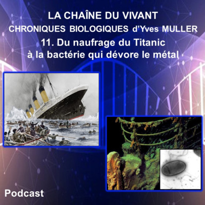
11. Du naufrage du Titanic à la bactérie qui dévore le métal
Cette 11ème chronique démarre le 10 avril 1912, avec le départ inaugural d’un immense paquebot transatlantique baptisé le Titanic et qui met le cap sur New-York. Mais dans la nuit du 14 au 15 avril 1912, le Titanic va malheureusement rencontrer un iceberg qui va provoquer son rapide naufrage. Cette chronique se poursuit par la découverte en 1985 de l’épave du Titanic, une carcasse métallique gisant à près de 4000 m de profondeur au large de Terre-Neuve. Cette épave est recouverte de sortes de stalactites de rouille. En 2010, une nouvelle bactérie est découverte dans des échantillons de ces stalactites de rouille récoltés auparavant sur l’épave du Titanic. Cette bactérie baptisée Halomonas titanicae est d’une part capable de supporter de grandes variations de salinité grâce à l’ectoïne qu’elle contient et elle est d’autre part responsable de l’accélération de la corrosion de la coque métallique du Titanic.
Auteur : Yves Muller – Professeur Agrégé de classe exceptionnelle – Docteur en Neurosciences - Université de Montpellier
11. Biological Chronicles - From the sinking of the Titanic to bacteria that devours metal
Our chronicle begins on April 10, 1912, with the maiden voyage of a huge transatlantic liner called the Titanic, which sets sail for New York. During the night of April 14-15, 1912, the Titanic unfortunately strikes an iceberg, causing it to sink rapidly. Our chronicle continues with the discovery in 1985 of the wreckage of the Titanic, a metal carcass lying nearly 4000 m deep off the coast of Newfoundland. The wreck is covered with a sort of rust stalactites or « rusticles ». In 2010, a new bacteria was discovered in samples of these rusticles previously collected from the wreck of the Titanic. This bacteria, known as Halomonas titanicae, is able to withstand large variations in salinity thanks to the ectoine it contains and is also responsible for the acceleration of corrosion of the Titanic's metal hull.
Références :
- Galinski E, Pfeiffer H et Trüper H (1985) 1,4,5,6-tetrahydro-2-methyl-4-pyrimidinecarboxylic acid. A novel cyclic amino acid from halophilic phototrophic bacteria of the genus Ectothiorhodospira - European Journal of Biochemistry ; 149:135-9
- Ballard R (1988) L'Exploration du Titanic - Glénat
- Gérard Piouffre (2009) Le Titanic ne répond plus - Larousse
- Sánchez-Porro C, Kaur B, Mann H et Ventosa A (2010) Halomonas titanicae sp. nov., a halophilic bacterium isolated from the RMS Titanic - International Journal of Systematic and Evolutionary Microbiology ; 12:2768-74
- Zaccai G, Bagyan I et coll (2016) Neutrons describe ectoine effects on water H-bonding and hydration around a soluble protein and a cell membrane - Scientific Reports ; 6:31434
- Dong Y, Lekbach Y et coll. (2020) Microbiologically influenced corrosion of 304L stainless steel caused by an alga associated bacterium Halomonas titanicae - Journal of Materials Science & Technology ; 37:200-6

10. Du Dieu grec Hermaphrodite au testicule féminisant
Cette 10ème chronique démarre avec un récit de la mythologie grecque écrit par le poète latin Ovide et qui nous raconte la rencontre entre le jeune Dieu Hermaphrodite et la belle nymphe Salmacis au creux de l’eau d’une source. Et Hermaphrodite va ressortir de cette source avec un corps à la fois masculin et féminin. La suite de cette chronique décrit dans le fœtus humain les étapes du processus de formation du système reproducteur masculin et féminin ainsi que les hormones impliquées. Puis sont abordées la maturation de ce système reproducteur et la mise en place des caractères sexuels secondaires qui ont lieu lors de la puberté chez le garçon et chez la fille. La chronique décrit ensuite les modes d’action de la testostérone et présente le récepteur des androgènes. En fin de chronique est décrit un syndrome étonnant présenté par certains sujets qui ont un génotype masculin et des testicules, mais dont les caractères phénotypiques sont féminins. Et nous verrons ainsi que la mutation d’un gène peut avoir des effets comparables à la source mythologique de Salmacis qui dévirilisa le jeune Hermaphrodite.
Auteur : Yves Muller – Professeur Agrégé de classe exceptionnelle - Docteur en Neurosciences - Université de Montpellier
10. Biological Chronicles - From the God Hermaphroditus to testicular feminization
This tenth chronicle begins with a story from Greek mythology written by the Latin poet Ovid which tells us about the meeting of the young God Hermaphrodite and the beautiful nymph Salmacis in the waters of a spring. Hermaphroditus emerges from the spring with a body that is both male and female. We follow this with the stages of formation of the male and female reproductive system in the human foetus as well as the hormones involved. Then we discuss the maturation of this reproductive system and the establishment of secondary sexual characteristics during puberty in boys and girls. Our chronicle then moves on to the modes of action of testosterone and presents the androgen receptor. Finally, we will discuss an astonishing syndrome found in certain subjects who have a male genotype and testicles, but whose phenotypic characters are female. And we will see that the mutation of a gene can have effects comparable to the mythological spring of Salmacis who emasculated the young Hermaphroditus.
Références :
- Ovide, Métamorphoses - Livre IV – (1er siècle après J.-C.) - Classiques- Le Livre de poche, édition de 2010
- Morris JM (1953) - The syndrome of testicular feminization in male pseudohermaphrodites - Am J Obstet Gynecol. - 65:1192-1211
- Brown TR (1995). "Human androgen insensitivity syndrome". Journal of Andrology. -16 (4): 299–303.
- Rachel A Davey and Mathis Grossmann (2016) - Androgen Receptor Structure, Function and Biology: From Bench to Bedside, Clin Biochem Rev. - 37(1):3–15
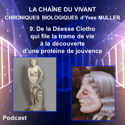
9. De la Déesse Clotho qui file la trame de vie à la découverte d’une protéine de jouvence
Cette 9ème chronique démarre avec 3 déesses de la mythologie grecque appelées les Moires et qui déterminent la destinée des hommes. Parmi ces 3 divinités, Clotho est la Moire qui tisse le fil de la vie des hommes. Cette chronique se poursuit par une surprenante découverte faite par des chercheurs japonais qui identifient en 1997 un gène dont la destruction dans une cellule œuf de souris provoque une forme spectaculaire de vieillissement prématuré. Et ces chercheurs vont donner à ce gène le nom de klotho. Depuis la découverte du gène klotho, de nombreuses études vont permettre de préciser les effets et le mode d’action de la protéine codée par ce gène. Klotho s’avère être non seulement une protéine de longévité, mais elle peut aussi favoriser le fonctionnement cérébral, retarder le développement des symptômes de maladies neurodégénératives et permettre de lutter contre certains cancers.
Auteur : Yves Muller – Professeur Agrégé de classe exceptionnelle - Docteur en Neurosciences - Université de Montpellier
9. Biological Chronicles - From the Goddess Clotho who spins the web of life, to the discovery of a protein of youth.
Our chronicle begins with 3 goddesses from Greek mythology called the Moirai, the Fates, who decide the destiny of men. Among these 3 divinities, Clotho is the Fate who weaves the thread of life. This chronicle continues with a surprising discovery made by Japanese researchers in 1997 who identified a gene whose destruction in the cell of a mouse egg caused a spectacular form of premature ageing. They gave the gene the name of klotho. Since the discovery of the klotho gene, numerous studies have been able to specify the effects and the mode of action of the protein encoded by this gene. Klotho has been shown to be not only a longevity protein, but it can also promote brain function, delay the development of symptoms of neurodegenerative diseases and help fight certain cancers.
Author : Yves Muller – Associate Professor at Montpellier University
Références :
- Jean Claude Ameisen - La sculpture du vivant – Le suicide cellulaire ou la mort créatrice – Editions du Seuil, 1999.
- Leon J, Moreno AJ, Garay BI, et coll. Peripheral elevation of a Klotho fragment enhances brain function and resilience in young, aging, and α-synuclein transgenic mice. Cell Reports 2017 Aug. 8, 20:1360-1371.
- Dubal DB, Zhu L, Sanchez PE, et coll. Life extension factor klotho prevents mortality and enhances cognition in hAPP transgenic mice. The Journal of Neuroscience 2015, 35:2358-71.
- Dubal DB, Yokoyama JS, Zhu L, et coll. Life extension factor klotho enhances cognition. Cell Reports 2014, 7:1065-76.
- Zeldich E, Chen CD, Colvin TA, et coll. The neuroprotective effect of Klotho is mediated via regulation of members of the redox system. The Journal of Biological Chemistry 2014, 289:24700-15.
- Kurosu H, Yamamoto M, Clark JD, et coll. Suppression of aging in mice by the hormone Klotho. Science 2005, 309:1829-33.
- Nagai T, Yamada K, Kim HC, et coll. Cognition impairment in the genetic model of aging klotho gene mutant mice: a role of oxidative stress. The FASEB Journal 2003, 17:50-2.
- Kuro-o M, Matsumura Y, Aizawa H, et coll. Mutation of the mouse klotho gene leads to a syndrome resembling ageing. Nature 1997, 390:45-51.
- Ella Zeldich, Ci-Di Chen, Emma Boden, et coll. Klotho Is Neuroprotective in the Superoxide Dismutase (SOD1G93A) Mouse Model of ALS. J Mol Neurosci 2019; 69 (2): 264-285.
- Jean Claude Ameisen, Clotho et le fil de vie, émission sur France Inter, Sur les épaules de Darwin (2017).
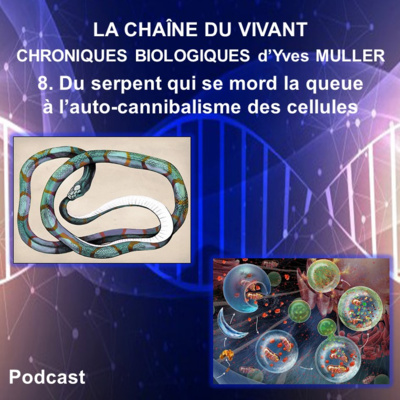
8. Du serpent qui se mord la queue à l’auto-cannibalisme des cellules
Cette 8ème chronique démarre avec l’un des plus anciens symboles mystiques du monde, appelé l’ouroboros, et qui est un serpent qui mord sa propre queue. Cette chronique aborde ainsi l’origine égyptienne de ce symbole, son utilisation par diverses civilisations puis ses différentes significations. Cette chronique décrit ensuite un étrange processus qui a lieu dans les cellules et au cours duquel une sorte de croissant limité par une membrane se forme, s’allonge et finit par se refermer comme un ouroboros. La fermeture de ce croissant membranaire aboutit à la formation d’une vacuole limitée par une double membrane et qu’on appelle l’autophagosome. Les lysosomes sont les organites de la digestion intracellulaire et ils vont permettre notamment la digestion du contenu de l’autophagosome. Et ainsi, comme le serpent qui dévore sa propre queue, les cellules peuvent digérer leurs propres constituants par un processus d’autophagie, une sorte d’auto-cannibalisme cellulaire. La formation d’un autophagosome caractérise la forme majeure d’autophagie appelée la macro-autophagie, mais cette chronique présente aussi d’autres types d’autophagie. Le rôle des gènes Atg (autophagy-related gene) dans l’autophagie est également évoqué. Puis cette chronique montre que l’autophagie joue un rôle essentiel à l’échelle des cellules et de l’organisme. Les relations entre l’autophagie et certaines maladies liées au vieillissement, telles que les cancers et les maladies neurodégénératives, sont également abordées. Et s’offre aujourd’hui à l’homme la possibilité de moduler l’autophagie pour lutter contre ces maladies. Dans la mythologie égyptienne, l’ouroboros protégeait le pharaon lors de son passage dans le monde des morts et lui permettait d’accéder à l’immortalité. L’autophagie de nos cellules nous protège aussi des méfaits du vieillissement et l’activer pourrait prolonger notre longévité.
Auteur : Yves Muller – Professeur Agrégé de classe exceptionnelle - Docteur en Neurosciences - Université de Montpellier
8. Biological Chronicles - From the snake that eats its tail to the auto-cannibalism of cells
This chronicle begins with one of the oldest mystical symbols in the world, the Ouroboros, the snake that eats its own tail. We thus addresses the Egyptian origin of this symbol, its use by various civilizations and then its different meanings. We then move on to a strange cellular process in which a kind of crescent surrounded by a membrane forms, lengthens and eventually closes in an ouroboros type membrane. The closure of this crescent membrane results in the formation of a vacuole limited by a double membrane called the autophagosome. Lysosomes are the organelles of intracellular digestion and which permit the digestion of the contents of the autophagosome. And so, like the snake that devours its own tail, cells can digest their own constituents through a process of autophagy, a kind of cellular auto-cannibalism. The formation of an autophagosome characterizes the major form of autophagy called macro-autophagy, but we will also present other types of autophagy. The role of the autophagy-related gene (Atg) in autophagy is also discussed. Then we will see how autophagy plays an essential role at the level of cells and the organism. The relationship between autophagy and certain age-related diseases, such as cancer and neurodegenerative diseases, is also discussed. Today humans have the possibility of modulating autophagy to fight against these diseases. In Egyptian mythology, the Ouroboros protected the pharaoh during his passage in the world of the dead and allowed him to reach immortality. The autophagy of our cells also protects us from the harms of ageing and activating it could prolong our longevity.
Author : Yves Muller – Associate Professor at Montpellier University
7. Du chant du canari à la régénération du cerveau
Cette 7ème chronique démarre par le chant de séduction qu’émet le canari mâle au printemps et que cet oiseau chanteur va modifier chaque année. Vous croiserez ensuite le Dieu Pan jouant de la flûte, des mésanges cachant des graines, des souris qui apprennent et mémorisent, des chauffeurs de taxi de la ville de Londres et des essais nucléaires pendant la guerre froide. Cette chronique vous conduira à découvrir que le cerveau des oiseaux et des mammifères adultes est pourvu d’une capacité de régénération. Et peut-être parviendrons-nous un jour à stimuler la production de neurones dans notre cerveau pour faire face à des lésions cérébrales ou des maladies neurodégénératives.
Auteur : Yves Muller - Professeur Agrégé de classe exceptionnelle - Docteur en Neurosciences - Université de Montpellier
7. Biological Chronicles - From the song of the canary to the regeneration of the brain
Our chronicle begins with the seduction song the male canary emits in spring and which this songbird will modify each year. We will then encounter the God Pan playing the flute, chickadees hiding seeds, mice with learning and memorizing capabilities, London taxi drivers and nuclear tests during the Cold War. Our chronicle leads us to then to the discovery that the brains of adult birds and mammals have a capacity for regeneration. And maybe we’ll one day be able to stimulate the production of neurons in our brain to deal with brain damage or neurodegenerative diseases. From a fly with legs on its head to the discovery architectural genes
Author : Yves Muller – Associate Professor at Montpellier University
Références :
- Messier B, Leblond CP, Smart I (1958) Presence of DNA synthesis and mitosis in the brain of young adult mice - Experimental Cell Research ; 14:224–6
- Altman J et Das GD (1965) Autoradiographic and histological evidence of postnatal hippocampal neurogenesis in rats - Journal of Comparative Neurology ; 124:319-35
- Nottebohm F, Stokes TM, Leonard CM (1976) Central control of song in the canary, Serinus canaria - Journal of Comparative Neurology ; 165: 457–86
- Nottebohm F (1981) A brain for all seasons: cyclical anatomical changes in song control nuclei of the canary brain - Science ; 214:1368-70
- Paton JA et Nottebohm FN (1984) Neurons generated in the adult brain are recruited into functional circuits – Science ; 225:1046-8
- Nottebohm F (1989) From Bird Song to Neurogenesis - Scientific American ; 260:74-9
- Alvarez-Buylla A, Theelen M et Nottebohm F (1990) Proliferation “hot spots” in adult avian ventricular zone reveal radial cell division – Neuron ; 5:101–9
- Kirn J, O’Loughlin B et coll. (1994) Cell death and neuronal recruitment in the high vocal center of adult male canaries are temporally related to changes in song - PNAS USA ; 91:7844-8
- Barena A et Nottebohm F (1994) Seasonal recruitment of hippocampal neurons in adult free-ranging black-capped chickadees - PNAS USA ; 91:11217-21
- Eriksson PS, Perfilieva E et coll. (1998) Neurogenesis in the adult human hippocampus - Nature Medicine ; 4:1313-7
- Taupin P (2005) Neurogenesis in the pathologies of the nervous system - Medecine Sciences 21:711-4
- Nottebohn F (2005) The neural basis of birdsong – PloS Biology ; 3:e164,759-61
- Rapanelli M, Frick L et Zanutto B (2011) Learning an operant conditioning task differentially induces gliogenesi in the medial prefrontal cortex and neurogenesis in the hippocampus - PLoS One 2011 ; 6:e14713, 1-12
- Woollett K et Maguire E (2011) Acquiring “the Knowledge” of London's layout drives structural brain changes - Current Biology ; 21:2109-14
- Bergmann O, Liebl J et coll. (2012) The age of olfactory bulb neurons in humans – Neuron ; 74:634-9
- Alonso M, Lepousez G et coll. (2012) Activation of adult-born neurons facilitates learning and memory - Nature Neuroscience ; 15:897-904

6. Du combat victorieux de David contre Goliath aux voies nerveuses de notre vision
Cette 6ème chronique biologique démarre par le célèbre combat décrit dans la Bible et qui vit David l'emporter face au gigantesque Goliath. Elle aborde ensuite l'origine du gigantisme, l'axe somatotrope, les rôles de l'hormone de croissance et l'acromégalie. Puis elle traite des voies nerveuses de la vision et les déficiences visuelles qui résultent de lésions de ces voies. Et cette chronique tentera finalement d'expliquer l'issue du combat de David contre Goliath à la lumière des connaissances médicales d'aujourd'hui.
Auteur : Yves Muller – Professeur Agrégé de classe exceptionnelle - Docteur en Neurosciences - Université de Montpellier
6. Biology Chronicles - From the victorious battle of David vs. Goliath to the nervous pathways of our sight.
This biological chronicle starts with the famous battle described in the Bible in which David defeated the gigantic Goliath. We will the discuss the origin of gigantism, the somatotropic axis, the roles of growth hormone and acromegaly. Then, we will with deal with nerve pathways of sight and vision, and the visual impairments that result from lesions on these pathways. Lastly, we will attempt to explain the outcome of David's fight against Goliath in light of today's medical knowledge.
Author : Yves Muller – Associate Professor at Montpellier University
Références :
- Gwilt JR. Biblical ills and remedies. Journal of the Royal Society of Medicine. 79, 1986, 738-742
- Sprecher S. David and Goliath Radiology. 1990 Jul;176(1):288.

5. D’un petit ver transparent aux gènes qui contrôlent le suicide de nos cellules
Cette 5ème chronique présente les mécanismes moléculaires impliqués dans une forme de mort cellulaire programmée qu’on appelle l’apoptose. Ces mécanismes sont d’abord explorés chez un petit ver transparent appelé Caenorhabditis elegans puis ils sont abordés chez les Mammifères. Sont ainsi présentés les caspases, les membres de la famille Bcl-2, l’Apaf-1, l’AIF, la voie extrinsèque de l’apoptose mettant en jeu les récepteurs de mort et la voie intrinsèque de l’apoptose mettant en jeu la mitochondrie et régulée par les membres de la famille Bcl-2. En allant ainsi du Nématode à l’homme, cette chronique permet de montrer que les mécanismes moléculaires qui conduisent nos cellules à subir l’apoptose ou au contraire à y résister ont été non seulement conservés au cours de l’évolution, mais qu’ils se sont aussi fortement diversifiés. La connaissance des ces mécanismes chez l’humain ouvre aussi de nouveaux espoirs dans la lutte contre certains cancers en utilisant des substances qui provoquent l’apoptose des cellules cancéreuses.
Auteur : Yves Muller – Professeur Agrégé de classe exceptionnelle - Docteur en Neurosciences - Université de Montpellier
5. Biological Chronicles - From a small transparent worm to the genes that control cellular suicide.
This chronicle presents the molecular mechanisms involved in a form of programmed cell death called apoptosis. We first explore these mechanisms in a small transparent worm called Caenorhabditis elegans and then in mammals. We will present Caspases, members of the Bcl-2 family, Apaf-1, AIF, the extrinsic pathway of apoptosis involving death receptors, and the participation of mitochondria in the intrinsic pathway of apoptosis and regulated by members of the Bcl-2 family. Thus going from the Nematode to man, this chronicle allows us to show that the molecular mechanisms that lead our cells to undergo apoptosis or, on the contrary, to resist it were not only preserved during evolution, but that they have also become highly diversified. The knowledge of these mechanisms in humans also opens new hopes in the fight against certain cancers by using substances that cause the apoptosis of cancer cells.
Author : Yves Muller – Associate Professor at Montpellier University
Références :
- Jean Claude Ameisen (1999) La sculpture du vivant – Le suicide cellulaire ou la mort créatrice – Editions du Seuil
-Susan Elmore (2007) Apoptosis: a review of programmed cell death » Toxicol. Pathol., vol. 35, no 4, p. 495-516
- N. Volkmann, F.M. Marassi, D.D. Newmeyer, D. Hanein (2014) The rheostat in the membrane: BCL-2 family proteins and apoptosis - Cell Death Differ., vol. 21, no 2, p. 206-15

4. Du chant des Sirènes à l’étude du suicide cellulaire chez un petit ver transparent
C’est le chant des Sirènes qui ouvre cette 4ème chronique biologique et ce chant envoûtant entraîne malheureusement ceux qui l’entendent vers la mort. Cette chronique évoque notamment les Sirènes de la mythologie grecque rencontrées par les Argonautes lors leur quête de la Toison d’Or. Et c’est la présence d’Orphée près des Argonautes qui permet à l’équipage d’échapper au chant mortel des Sirènes parce que le chant d’Orphée neutralise celui des Sirènes. Cette chronique présente ensuite un petit ver transparent appelé Caenorhabditis elegans et dont le développement embryonnaire met en jeu la mort programmée de certaines cellules embryonnaires par un processus appelé l’apoptose. Et l’étude d’embryons porteurs de mutations génétiques va montrer que la survie ou l’apoptose de chaque cellule embryonnaire dépend de la présence de 4 gènes, ced-3, ced-4, egl-1 et ced-9, codant 4 protéines essentielles. Les protéines CED-3, CED4 et EGL-1 sont comme des sirènes présentes dans les cellules de l’embryon et conduisant à l’apoptose alors que la protéine CED-9 est, comme Orphée, un protecteur neutralisant le chant mort des Sirènes et permettant donc aux cellules de l’embryon de réprimer leur autodestruction.
Auteur : Yves Muller – Professeur Agrégé de classe exceptionnelle - Docteur en Neurosciences - Université de Montpellier
4. Biological Chronicles - From the siren song to the study of cell suicide in a small transparent worm
We open this biological chronicle with the song of Sirens which, sadly, brings death to all those who hear it . This chronicle evokes the Sirens of Greek mythology encountered by the Argonauts during their quest for the Golden Fleece. It’s thanks to the presence of Orpheus near the Argonauts that the crew are able to escape from the mortal song. Orpheus’ song neutralizes that of the Sirens. This chronicle then presents a small transparent worm called Caenorhabditis elegans whose embryonic development involves the programmed death of certain embryonic cells by a process called apoptosis. The study of embryos carrying genetic mutations will show that the survival or apoptosis of each embryonic cell depends on the presence of 4 genes, ced-3, ced-4, egl-1 and ced-9, coding 4 essential proteins. The CED-3, CED4 and EGL-1 proteins are like sirens present in the cells of the embryo and lead to apoptosis. The CED-9 protein is, like Orpheus, a neutralizing protector for the embryonic cells to suppress their self-destruction.
Author : Yves Muller – Associate Professor at Montpellier University
Références :
- Jean Claude Ameisen (1999) La sculpture du vivant – Le suicide cellulaire ou la mort créatrice – Editions du Seuil
- Barbara Conradt, Yi-Chun Wu and View ORCID ProfileDing Xue (2016) Programmed Cell Death During Caenorhabditis elegans Development - Genetics vol. 203 no. 4 1533-1562

3. De Pénélope tissant sa toile à l’enzyme qui efface le temps
Cette 3ème chronique démarre avec un récit de la mythologie grecque présent dans le poème « L’Odyssée » écrit par le poète Homère. Ce récit nous raconte comment la fidèle Pénélope parvient à repousser ses prétendants en attendant durant 20 ans le retour de son mari Ulysse. Elle a promis à ses soupirants de choisir l’un d’entre eux le jour où elle finira de tisser le linceul destiné au père d’Ulysse. Mais elle va défaire chaque nuit la toile qu’elle tisse le jour, parvenant par ce subterfuge à suspendre le temps. Puis cette chronique se poursuit par une étonnante histoire scientifique qui va nous conduire à la découverte d’une enzyme appelée la télomérase. Cette enzyme reconstruit dans certaines de nos cellules les parties terminales de nos chromosomes appelées les télomères, et que les générations cellulaires successives tendent normalement à raccourcir. Et nous verrons que la télomérase, comme Pénélope, permet de suspendre le temps.
Auteur : Yves Muller – Professeur Agrégé de classe exceptionnelle - Docteur en Neurosciences - Université de Montpellier
3. Biological Chronicles - From Penelope weaving her shroud to the enzyme that erases time
Our chronicle begins with an account of Greek mythology from "The Odyssey" written by the poet Homer. This story tells us how the faithful Penelope manages to hold off her suitors for twenty years while awaiting the return of her husband Ulysses. She promises her suitors that she will make her choice the day she finishes weaving the shroud for Ulysses' father. But each night she undoes the canvas woven during the day, thus managing to suspend time. Our chronicle continues with an astonishing scientific story which leads us to the discovery of an enzyme called telomerase. In certain cells, this enzyme rebuilds the terminal parts of our chromosomes called telomeres, which successive cell generations normally tend to shorten. And we will see that telomerase, like Penelope, makes it possible to suspend time.
Références :
- Baker D, Childs B, Durik M, et coll. Naturally occurring p16(Ink4a)-positive cells shorten healthy lifespan. Nature 2016, 530:184-9.
- Dilley R, Verma P, Cho N, et coll. Break-induced telomere synthesis underlies alternative telomere maintenance. Nature 2016, 539:54-8.
- Blackburn E, Epel E, Lin J. Human telomere biology: A contributory and interactive factor in aging, disease risks, and protection. Science 2015, 350:1193-8.
- Van Deursen J. The role of senescent cells in ageing. Nature 2014, 509:439-46.
- Olovnikov A. Telomeres, telomerase, and aging: origin of the theory. Experimental Gerontology 1996, 31:443-8.
- Yu G, Bradley J, Attardi L, Blackburn E. In vivo alteration of telomere sequences and senescence caused by mutated Tetrahymena telomerase RNAs. Nature 1990, 344:126‐32.
- Lundblad V, Szostak J. A mutant with a defect in telomere elongation leads to senescence in yeast. Cell 1989, 57:633‐43.
- Greider C, Blackburn E. A telomeric sequence in the RNA of Tetrahymena telomerase required for telomere repeat synthesis. Nature 1989, 337:331‐7.
- Greider C, Blackburn E. The telomere terminal transferase of Tetrahymena is a ribonucleoprotein enzyme with two kinds of primer specificity. Cell1987, 51:887‐98.
- Greider C, Blackburn E. Identification of a specific telomere terminal transferase activity in Tetrahymena extracts. Cell 1985, 43:405‐13.
- Szostak J, Blackburn E. Cloning yeast telomeres on linear plasmid vectors. Cell1982, 29:245‐55.
- Olovnikov A. A theory of marginotomy. Journal of Theoretical Biology 1973, 41:181-90.
- Hayflick L, Moorhead P. The serial cultivation of human diploid cell strains. Experimental Cell Research 1961, 25:585-621.

2. Du Parc National de Yellowstone à la photocopieuse à gènes
Cette 2ème chronique biologique démarre par l’exploration du Parc National de Yellowstone aux États-Unis, un parc réputé notamment pour son intense activité géothermique. Dans les années 1960, un biologiste va découvrir dans une des sources chaudes de Yellowstone une nouvelle bactérie capable de vivre dans des eaux pouvant atteindre 80°C. La découverte de cette bactérie, appelée Thermus aquaticus, va ouvrir le champ d’étude des organismes capables de vivre dans des milieux extrêmes. Mais cette découverte va aussi conduire à une révolution en biologie moléculaire grâce à l’utilisation de l’ADN polymérase de cette bactérie dans la technique d’amplification de l’ADN appelée la PCR (Polymerase Chain Reaction).
Auteur : Yves Muller – Professeur Agrégé de classe exceptionnelle - Docteur en Neurosciences - Université de Montpellier
2. Biology Chronicles - From Yellowstone National Park to the Gene Copying Machine
Our biological chronicle starts with the exploration of Yellowstone National Park in the USA, a park renowned for its intense geothermal activity. In the 1960s, in one of the hot springs of Yellowstone a new bacterium was discovered by a biologist that is capable of living in waters up to 80 ° C. The discovery of this bacterium, called Thermus aquaticus, opened the field of study of organisms capable of living in extreme environments. But this discovery also led to a revolution in molecular biology through the use of the DNA polymerase of this bacterium in the DNA amplification technique called PCR (Polymerase Chain Reaction).
Author : Yves Muller – Associate Professor at Montpellier University
Références :
Chien A, Edgar DB, Trela JM (1976). "Deoxyribonucleic acid polymerase from the extreme thermophile Thermus aquaticus". Journal of Bacteriology. 127 (3): 1550–7.
Mullis KB (April 1990). "The unusual origin of the polymerase chain reaction". Scientific American. 262 (4): 56–61, 64–5.
Ishino S, Ishino Y (2014). "DNA polymerases as useful reagents for biotechnology - the history of developmental research in the field". Frontiers in Microbiology. 5: 465.
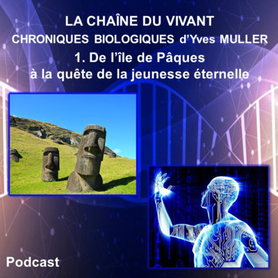
1. De l’île de Pâques à la quête de la jeunesse éternelle
Cette 1ère chronique biologique vous conduira d’une bactérie trouvée dans le sol de l’île de Pâques à la découverte d’une molécule capable de prolonger la vie des animaux.
Auteur : Yves Muller – Professeur Agrégé de classe exceptionnelle - Docteur en Neurosciences - Université de Montpellier
1. Biological Chronicles - From Easter Island to search of eternal youth
It’s 1964, and a group of Canadian scientists had sailed across the Pacific to Easter Island in order to study the health of the isolated local population. Working below the gaze of the island’s famous statues, they collected a variety of soil samples and other biological material, unaware that one of these would yield an unexpected treasure. It contained a bacterium that secreted a new antibiotic, one that proved to be a potent anti-fungal chemical. The compound was named rapamycin after the traditional name of its island source – Rapa Nui. Rapamycin has made a stunning journey from the soil of a Pacific island to the besides of the world’s hospitals. Its ability to suppress the immune system means that it’s given to transplant patients to stop them from rejecting their organs and its ability to stop cells from dividing has formed the basis of potential anti-cancer drugs. But the chemical has an even more interesting ability and one that has only just been discovered – it can extend lifespan, for example in mice.